Renal_oncocytoma4.jpg

Size of this preview:
729 × 599 pixels
.
Other resolutions:
292 × 240 pixels
|
584 × 480 pixels
|
934 × 768 pixels
|
1,245 × 1,024 pixels
|
1,868 × 1,536 pixels
.
Summary
| Description |
English:
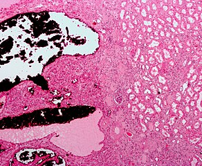
Low magnification
micrograph
of a
renal oncocytoma
.
H&E stain
.
The tumour cells (left of the image) are arranged in nests, have slightly enlarged nuclei and have a more eosinophilic (darker pink) cytoplasm than the normal kidney (right of image). A glomerulus is seen at the centre of the image. Normal renal tubules are seen on the right of the image. On electronmicroscopy , oncocytomas have abundant mitochondria . See alsoImage:Renal_oncocytoma2.jpg - high magnification. Image:Renal_oncocytoma3.jpg - intermediate magnification. |
| Date | |
| Source | Own work |
| Author | Nephron |
Licensing
I, the copyright holder of this work, hereby publish it under the following licenses:
This file is licensed under the
Creative Commons
Attribution-Share Alike 3.0 Unported
license.
-
You are free:
- to share – to copy, distribute and transmit the work
- to remix – to adapt the work
-
Under the following conditions:
- attribution – You must give appropriate credit, provide a link to the license, and indicate if changes were made. You may do so in any reasonable manner, but not in any way that suggests the licensor endorses you or your use.
- share alike – If you remix, transform, or build upon the material, you must distribute your contributions under the same or compatible license as the original.

|
Permission is granted to copy, distribute and/or modify this document under the terms of the GNU Free Documentation License , Version 1.2 or any later version published by the Free Software Foundation ; with no Invariant Sections, no Front-Cover Texts, and no Back-Cover Texts. A copy of the license is included in the section entitled GNU Free Documentation License . |
You may select the license of your choice.