Parasite140120-fig3_Acanthamoeba_keratitis_Figure_3B.png
Size of this preview:
580 × 600 pixels
.
Other resolutions:
232 × 240 pixels
|
464 × 480 pixels
|
742 × 768 pixels
|
990 × 1,024 pixels
|
2,032 × 2,102 pixels
.
Summary
| Description |
English:
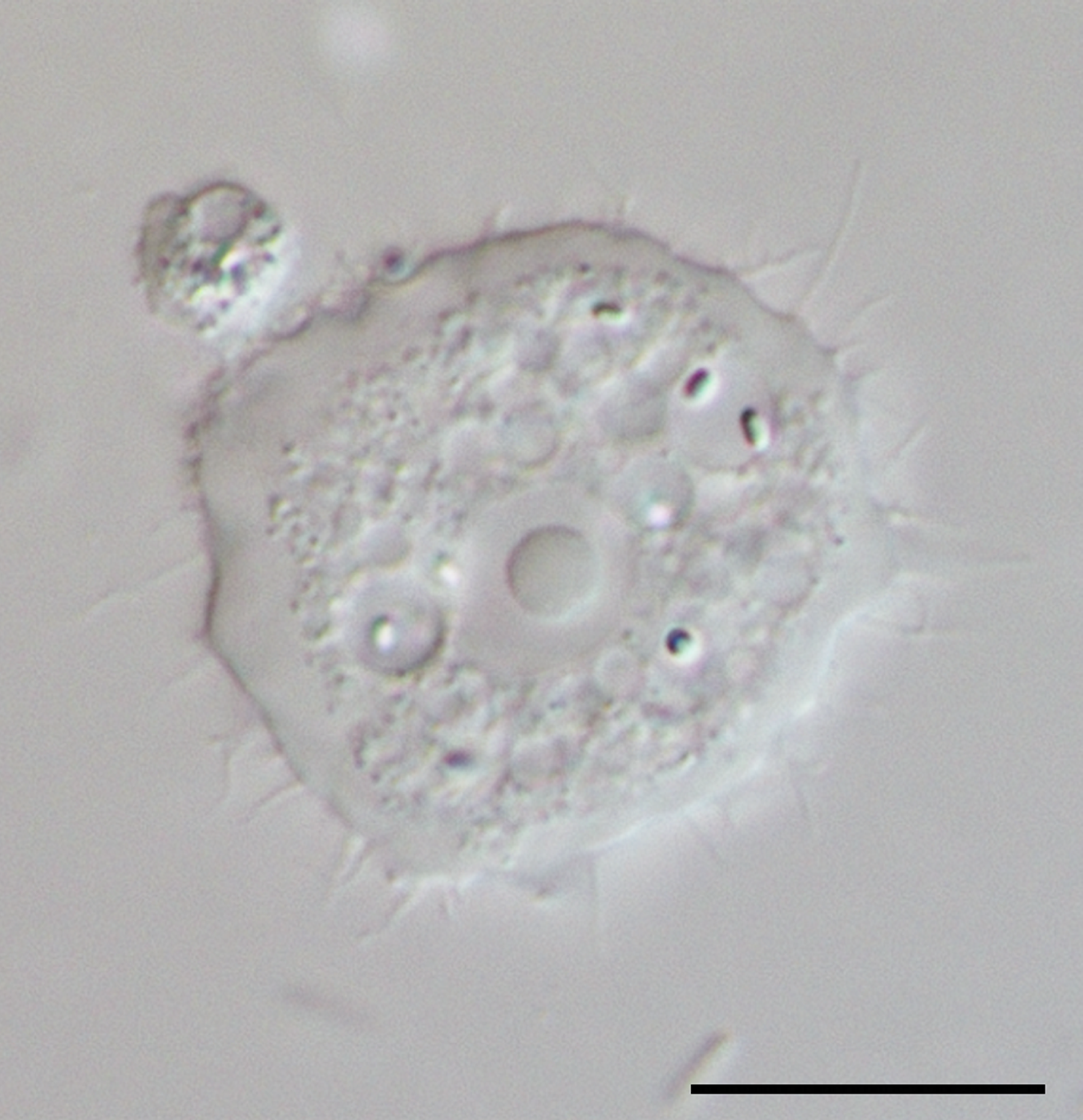
Figure 3B
of published paper.
Acanthamoeba trophozoites with the characteristic acanthopodia in bright field microscopy. Scale bar: 10 μm. Originals. |
| Date | |
| Source | " (2015). " An update on Acanthamoeba keratitis: diagnosis, pathogenesis and treatment ". Parasite 22 : 10. DOI : 10.1051/parasite/2015010 . PMID 25687209 . ISSN 1776-1042 . |
| Author | Jacob Lorenzo-Morales, Naveed A. Khan and Julia Walochnik |

Licensing
|
This file is licensed under the
Creative Commons
Attribution 4.0 International
license.
|

This file was published in the scientific journal
Parasite
.
Their website
states that all content of the journal including and after 2013 is published under the Creative Commons Attribution 4.0 license.
|