Microvilli.jpg
No higher resolution available.
Summary
| Description |
English:
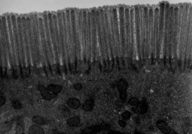
Transmission electron microscope image of a thin section cut through a human jejunum (segment of small intestine) epithelial cell. Image shows apical end of absorptive cell with some of the densely packed microvilli that make up the striated border. Each microvillus is approximately 1um long by 0.1um in diameter and contains a core of actin microfilaments.
|
| Source | http://remf.dartmouth.edu/images/humanMicrovilliTEM/source/1.html |
| Author | Louisa Howard, Katherine Connollly - Dartmouth Electron Microscope Facility |
|
Permission
( Reusing this file ) |
http://remf.dartmouth.edu/imagesindex.html |
Licensing
|
|
This work has been released into the
public domain
by its author,
Dartmouth Electron Microscope Facility
. This applies worldwide.
In some countries this may not be legally possible; if so: Dartmouth Electron Microscope Facility grants anyone the right to use this work for any purpose , without any conditions, unless such conditions are required by law.
|
|
|
This file was reviewed on 18:28, 13 October 2011 (UTC) by the
administrator
or
trusted user
Common Good
(
talk
)
, who confirmed the
Public Domain
status on that date.
|
Original upload log
Originally from en.wikipedia ; description page is (was) here
- 18:51, 28 December 2002 Magnus Manske 350x247 (19,689 bytes) (Source and public domain notice at [http://remf.dartmouth.edu/imagesindex.html])