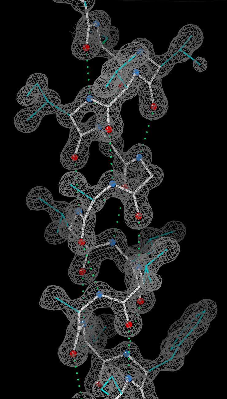
Helix_electron_density_myoglobin_2nrl_17-32.jpg
Size of this preview:
341 × 599 pixels
.
Other resolutions:
136 × 240 pixels
|
273 × 480 pixels
|
740 × 1,300 pixels
.
Summary
| Description |
English:
An alpha-helix, with stick-figures for the model shown within electron density for the crystal structure at ultra-high-resolution (0.91Å). The density contours are in gray, the helix backbone in white, sidechains in cyan, O atoms in red, N atoms in blue, and hydrogen bonds as green dotted lines. From PDB file 2NRL, residues 17-32.
|
| Date | |
| Source | Own work |
| Author | Dcrjsr |
Licensing
This file is licensed under the
Creative Commons
Attribution 3.0 Unported
license.
-
You are free:
- to share – to copy, distribute and transmit the work
- to remix – to adapt the work
-
Under the following conditions:
- attribution – You must give appropriate credit, provide a link to the license, and indicate if changes were made. You may do so in any reasonable manner, but not in any way that suggests the licensor endorses you or your use.