Gorham's_disease.jpg
No higher resolution available.
| Description |
English:
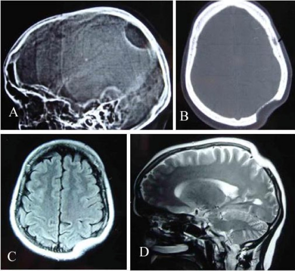
X-ray skull lateral view (A) showing a osteolytic area in left parietal region. CT scan bony window (B), MRI T1W Axial (C) and T2W Sagittal (D) revealing skull defect with normal brain parenchyma.
|
| Date | |
| Source | Parihar V, Yadav YR, Sharma D. Gorham's disease involving the left parietal bone: a case report Cases J. 1. 1, s. 258 (2008). doi:10.1186/1757-1626-1-258. PMID 18940015 . |
| Author | see above |
|
Permission
( Reusing this file ) |
http://www.biomedcentral.com/info/about/license |
This file is licensed under the
Creative Commons
Attribution 2.0 Generic
license.
-
You are free:
- to share – to copy, distribute and transmit the work
- to remix – to adapt the work
-
Under the following conditions:
- attribution – You must give appropriate credit, provide a link to the license, and indicate if changes were made. You may do so in any reasonable manner, but not in any way that suggests the licensor endorses you or your use.