Fusiform_face_area_face_recognition.jpg
No higher resolution available.
Summary
| Description |
English:
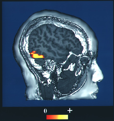
This is a computer-enhanced fMRI scan of a person who has been asked to look at faces. The image shows increased blood flow in the part of the visual cortex that recognizes faces.
日本語:
fMRI。顔を見るように言われた人の脳内で、視覚皮質の顔情報を処理する部位で、血流増加が起きている、という画像。
|
| Date | upload to commons at 2009-10-27 |
| Source | https://www.nlm.nih.gov/hmd/emotions/frontiers.html ( archive.org ) |
| Author | NIH |
|
Permission
( Reusing this file ) |
Public domain US government |
|
|
This image is a work of the
National Institutes of Health
, part of the
United States Department of Health and Human Services
, taken or made as part of an employee's official duties. As a
work
of the
U.S. federal government
, the image is in the
public domain
.

|
|
| This file has been identified as being free of known restrictions under copyright law, including all related and neighboring rights. | ||