CT_brain_tumor.jpg
Size of this preview:
499 × 599 pixels
.
Other resolutions:
200 × 240 pixels
|
400 × 480 pixels
|
640 × 768 pixels
|
853 × 1,024 pixels
|
1,437 × 1,725 pixels
.
Summary
| Description |
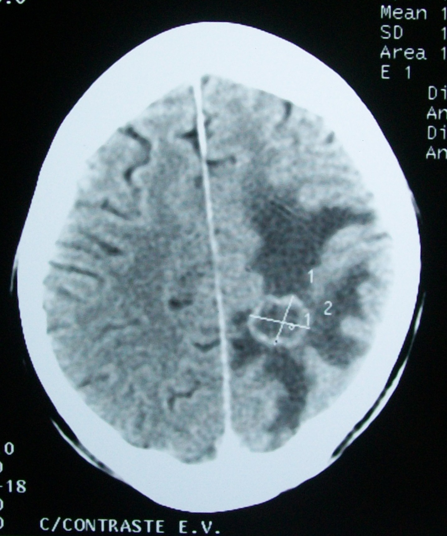
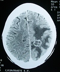
English:
CT scan
of a
metastasis
-suspected space-occupying
brain tumor
in the left temporo-parietal. There is hypoattenuating (dark)
peritumoral edema
in the surrounding white matter, with a "finger-like" spread. Same lesion seen by
MRI
:
File:MRI brain tumor.jpg
.
Español:
Lesion ocupante de espacio compatible con infiltración secundaria (
metástasis
) a nivel temporoparietal izquierda asociado con
edema
alrededor de la lesión.
TAC de cráneo
; misma lesión por TAC por
Resonancia magnética
de cráneo:
File:MRI brain tumor.jpg
.
|
| Date | |
| Source | Own work |
| Author | Bobjgalindo |
Licensing
I, the copyright holder of this work, hereby publish it under the following licenses:

|
Permission is granted to copy, distribute and/or modify this document under the terms of the GNU Free Documentation License , Version 1.2 or any later version published by the Free Software Foundation ; with no Invariant Sections, no Front-Cover Texts, and no Back-Cover Texts. A copy of the license is included in the section entitled GNU Free Documentation License . |
This file is licensed under the
Creative Commons
Attribution-Share Alike
Attribution-Share Alike 4.0 International
,
3.0 Unported
,
2.5 Generic
,
2.0 Generic
and
1.0 Generic
license.
-
You are free:
- to share – to copy, distribute and transmit the work
- to remix – to adapt the work
-
Under the following conditions:
- attribution – You must give appropriate credit, provide a link to the license, and indicate if changes were made. You may do so in any reasonable manner, but not in any way that suggests the licensor endorses you or your use.
- share alike – If you remix, transform, or build upon the material, you must distribute your contributions under the same or compatible license as the original.
You may select the license of your choice.