CADASIL.jpg
Size of this preview:
632 × 600 pixels
.
Other resolutions:
253 × 240 pixels
|
506 × 480 pixels
|
809 × 768 pixels
|
1,200 × 1,139 pixels
.
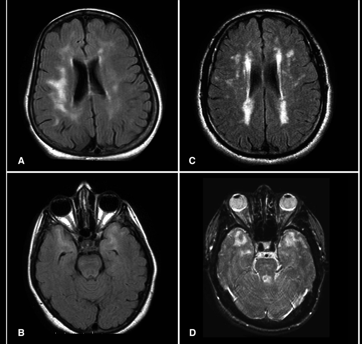
| Description | Axial FLAIR (a, b & c) and T2 weighted (d) Brain MRI from patients with CADASIL. The exams in 2a and 2b are from asymptomatic patients with depression. Note temporal lobe lesions even in asymptomatic patients (2b). In Figure 2a and d periventricular diffuse white matter ischemic lesion and multiple lacunar lesions in thalamus, pons and basal ganglia |
| Date | |
| Source | CADASIL in Arabs: clinical and genetic findings. BMC Medical Genetics 2007, 8:67doi:10.1186/1471-2350-8-67 |
| Author | Bohlega S, Al Shubili A, Edris A, Alreshaid A, Alkhairallah T, AlSous MW, Farah S, Abu-Amero KK. |
This file is licensed under the
Creative Commons
Attribution 2.0 Generic
license.
-
You are free:
- to share – to copy, distribute and transmit the work
- to remix – to adapt the work
-
Under the following conditions:
- attribution – You must give appropriate credit, provide a link to the license, and indicate if changes were made. You may do so in any reasonable manner, but not in any way that suggests the licensor endorses you or your use.