Anterior_mediastinal_mass_thymoma_diagram.jpg
Size of this preview:
800 × 599 pixels
.
Other resolutions:
320 × 239 pixels
|
640 × 479 pixels
|
858 × 642 pixels
.
Summary
| Description |
English:
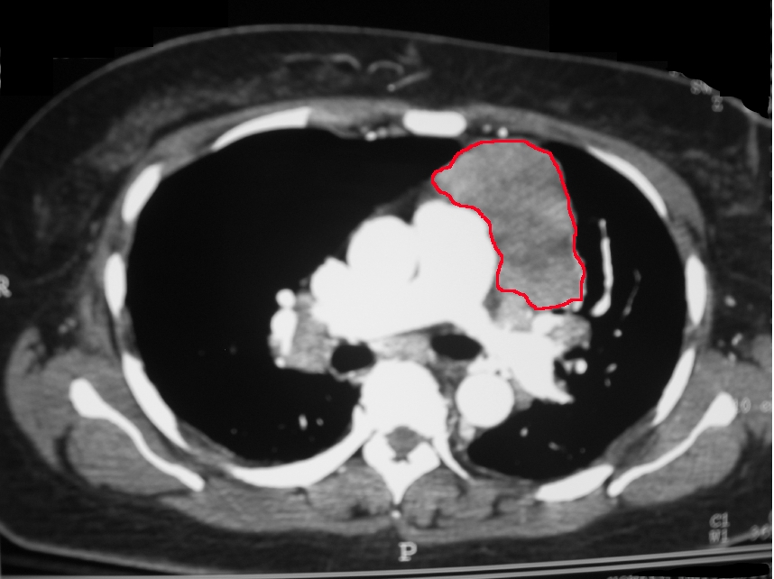
CT scan of the chest revealing a large necrotic mass in the left anterior mediastinum (later proven to be a thymoma) and bilateral hilar lymphadenopathy (from concurrent sarcoidosis).
|
| Date | Published: 25 July 2008 |
| Source | Rare association of thymoma, myasthenia gravis and sarcoidosis : a case report . Journal of Medical Case Reports . 2008; 2 :245. doi:10.1186/1752-1947-2-245 |
| Author | Mohankumar Kurukumbi, Roger L Weir, Janaki Kalyanam, Mansoor Nasim, Annapurni Jayam-Trouth. |
|
|
This is a
retouched picture
, which means that it has been digitally altered from its original version. Modifications:
Added red line indicating the tumor
. The original can be viewed here:
Anterior mediastinal mass thymoma.jpg
:
|
Licensing
This file is licensed under the
Creative Commons
Attribution 2.0 Generic
license.
-
You are free:
- to share – to copy, distribute and transmit the work
- to remix – to adapt the work
-
Under the following conditions:
- attribution – You must give appropriate credit, provide a link to the license, and indicate if changes were made. You may do so in any reasonable manner, but not in any way that suggests the licensor endorses you or your use.