Vacuolar_interface_dermatitis

Vacuolar interface dermatitis

Vacuolar interface dermatitis

Add article description


Vacuolar interface dermatitis (VAC, also known as liquefaction degeneration, vacuolar alteration or hydropic degeneration) is a dermatitis with vacuolization at the dermoepidermal junction, with lymphocytic inflammation at the epidermis and dermis.[1]

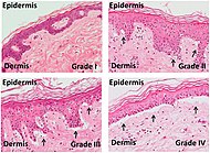
Vacuolar interface dermatitis, with lymphocytes in the dermis and epidermis (black arrow indicates one), and vacuolization (white arrow) at the dermoepidermal junction.
Micrograph of a vacuolar interface dermatitis with dermal mucin, as may be seen in lupus. H&E stain.

Causes

More information Main conditions, Characteristics ...

An interface dermatitis with vacuolar alteration, not otherwise specified, may be caused by viral exanthems, phototoxic dermatitis, acute radiation dermatitis, erythema dyschromicum perstans, lupus erythematosus and dermatomyositis.[2]


References

  1. Bolognia, Jean L.; et al. (2007). Dermatology. St. Louis: Mosby. p. 11. ISBN 1-4160-2999-0.
  2. Unless else specified in boxes, reference is: Alsaad, K O (2005). "My approach to superficial inflammatory dermatoses". Journal of Clinical Pathology. 58 (12): 1233–1241. doi:10.1136/jcp.2005.027151. ISSN 0021-9746.
  3. Lisa K Pappas-Taffer. "Lichen Sclerosus". Medscape. Updated: May 17, 2018
  4. Mowafak Hamodat. "Skin inflammatory (nontumor) > Lichenoid and interface reaction patterns > Lupus: systemic lupus erythematosus (SLE)". PathologyOutlines. Topic Completed: 1 August 2011. Revised: 26 March 2019

Share this article:

This article uses material from the Wikipedia article Vacuolar_interface_dermatitis, and is written by contributors. Text is available under a CC BY-SA 4.0 International License; additional terms may apply. Images, videos and audio are available under their respective licenses.