Phycobiliprotein
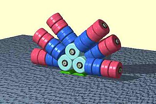
Phycobiliproteins are water-soluble proteins present in cyanobacteria and certain algae (rhodophytes, cryptomonads, glaucocystophytes). They capture light energy, which is then passed on to chlorophylls during photosynthesis. Phycobiliproteins are formed of a complex between proteins and covalently bound phycobilins that act as chromophores (the light-capturing part). They are most important constituents of the phycobilisomes.

This article needs additional citations for verification. (December 2009) |
| Phycobiliprotein | MW (kDa) | Ex (nm) / Em (nm) | Quantum yield | Molar Extinction Coefficient (M−1cm−1) | Comment | Image |
|---|---|---|---|---|---|---|
| R-Phycoerythrin (R-PE) | 240 | 498.546.566 nm / 576 nm | 0,84 | 1.53 106 | Can be excited by Kr/Ar laser Applications for R-Phycoerythrin Many applications and instruments were developed specifically for R-phycoerythrin. It is commonly used in immunoassays such as FACS, flow cytometry, multimer/tetramer applications. Structural Characteristics R-phycoerythrin is also produced by certain red algae. The protein is made up of at least three different subunits and varies according to the species of algae that produces it. The subunit structure of the most common R-PE is (αβ)6γ. The α subunit has two phycoerythrobilins (PEB), the β subunit has 2 or 3 PEBs and one phycourobilin (PUB), while the different gamma subunits are reported to have 3 PEB and 2 PUB (γ1) or 1 or 2 PEB and 1 PUB (γ2). |
 |
| B-Phycoerythrin (B-PE) | 240 | 546.566 nm / 576 nm | 0,98 | (545 nm) 2.4 106
(563 nm) 2.33 106 |
Applications for B-Phycoerythrin
Because of its high quantum yield, B-PE is considered the world's brightest fluorophore. It is compatible with commonly available lasers and gives exceptional results in flow cytometry, Luminex and immunofluorescent staining. B-PE is also less "sticky" than common synthetic fluorophores and therefore gives less background interference. Structural Characteristics B-phycoerythrin (B-PE) is produced by certain red algae such as Rhodella sp. The specific spectral characteristics are a result of the composition of its subunits. B-PE is composed of at least three subunits and sometimes more. The chromophore distribution is as follows: α subunit with 2 phycoerythrobilins (PEB), β subunit with 3 PEB, and the γ subunit with 2 PEB and 2 phycourobilins (PUB). The quaternary structure is reported as (αβ)6γ. |
 |
| C-Phycocyanin (CPC) | 232 | 620 nm / 642 nm | 0,81 | 1.54 106 | Accepts the fluorescence for R-PE; Its red fluorescence can be transmitted to Allophycocyanin | |
| Allophycocyanin (APC) | 105 | 651 nm / 662 nm | 0,68 | 7.3 105 | Excited by He/Ne laser; double labeling with Sulfo-Rhodamine 101 or any other equivalent fluorochrome. Applications for Allophycocyanin Many applications and instruments were developed specifically for allophycocyanin. It is commonly used in immunoassays such as flow cytometry and high-throughput screening. It is also a common acceptor dye for FRET assays. Structural Characteristics Allophycocyanin can be isolated from various species of red or blue-green algae, each producing slightly different forms of the molecule. It is composed of two different subunits (α and β) in which each subunit has one phycocyanobilin (PCB) chromophore. The subunit structure for APC has been determined as (αβ)3. |
 |
| ↑ = FluoProbes PhycoBiliProteins data | ||||||
Phycobiliproteins demonstrate superior fluorescent properties compared to small organic fluorophores, especially when high sensitivity or multicolor detection required :
- Broad and high absorption of light suits many light sources
- Very intense emission of light: 10-20 times brighter than small organic fluorophores
- Relative large Stokes shift gives low background, and allows multicolor detections.
- Excitation and emission spectra do not overlap compared to conventional organic dyes.
- Can be used in tandem (simultaneous use by FRET) with conventional chromophores (i.e. PE and FITC, or APC and SR101 with the same light source).
- Longer fluorescence retention period.
- High water solubility
Phycobiliproteins allow very high detection sensitivity, and can be used in various fluorescence based techniques fluorimetric microplate assays Archived 2018-03-18 at the Wayback Machine,[6][7][8] FISH and multicolor detection.
They are under development for use in artificial photosynthesis, limited by the relatively low conversion efficiency of 4-5%.[9]
- Contreras-Martel, C.; Legrand, P.; Piras, C.; Vernede, X.; et al. (2000-05-09). "Crystal structure of R-phycoerythrin at 2.2 angstroms". RCSB Protein Data Bank (PDB). doi:10.2210/pdb1eyx/pdb. PDB ID: 1EYX. Retrieved 11 October 2012.
{{cite journal}}: Cite journal requires|journal=(help) - Contreras-Martel C, Martinez-Oyanedel J, Bunster M, Legrand P, Piras C, Vernede X, Fontecilla-Camps JC (January 2001). "Crystallization and 2.2 A resolution structure of R-phycoerythrin from Gracilaria chilensis: a case of perfect hemihedral twinning". Acta Crystallographica D. 57 (Pt 1): 52–60. doi:10.1107/S0907444900015274. PMID 11134927. S2CID 216930. PDB ID: 1EYX.
- Camara-Artigas, A. (2011-12-16). "Crystal Structure of the B-phycoerythrin from the red algae Porphyridium cruentum at pH8". RCSB Protein Data Bank (PDB). doi:10.2210/pdb3v57/pdb. PDB ID: 3V57. Retrieved 12 October 2012.
{{cite journal}}: Cite journal requires|journal=(help) - Camara-Artigas A, Bacarizo J, Andujar-Sanchez M, Ortiz-Salmeron E, Mesa-Valle C, Cuadri C, Martin-Garcia JM, Martinez-Rodriguez S, Mazzuca-Sobczuk T, Ibañez MJ, Allen JP (October 2012). "pH-dependent structural conformations of B-phycoerythrin from Porphyridium cruentum". The FEBS Journal. 279 (19): 3680–3691. doi:10.1111/j.1742-4658.2012.08730.x. PMID 22863205. S2CID 31253970. PDB ID: 3V57.
- "Flow Cytometry" (PDF). Archived from the original (PDF) on 2018-03-18. Retrieved 2014-06-07.
- Telford, William G; Moss, Mark W; Morseman, John P; Allnutt, F.C.Thomas (August 2001). "Cyanobacterial stabilized phycobilisomes as fluorochromes for extracellular antigen detection by flow cytometry". Journal of Immunological Methods. 254 (1–2): 13–30. doi:10.1016/s0022-1759(01)00367-2. ISSN 0022-1759. PMID 11406150.
- Lavars, Nick (2021-10-19). "Encasing algae triples the efficiency of artificial photosynthesis". New Atlas. Retrieved 2021-10-24.