Coronavirus._SARS-CoV-2.png

Summary
| Description |
Deutsch:
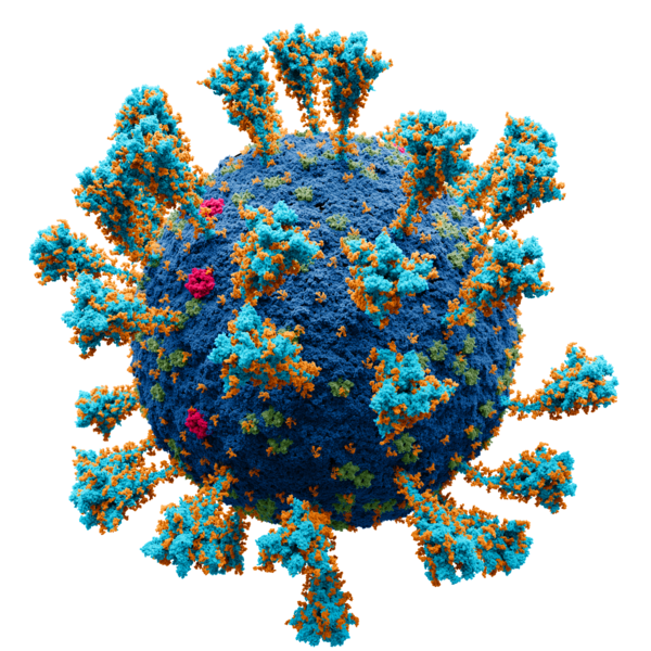
Wissenschaftlich genaues Atommodell der äußeren Struktur des
SARS
Coronavirus 2 (
SARS-CoV-2
), einem
Stamm
(genetische Variante) des Coronavirus, der die Coronavirus-Krankheit (
COVID-19
) verursachte und erstmals im Dezember 2019 in
Wuhan
,
China
, identifiziert wurde.
Jeder einzelne Ort (amorpher Fleck) ist ein Atom von:
kobalt:
Virushülle
purpurrot:
Hüllproteine
grün:
Matrixproteine
türkis:
Spike-Proteine
English:
Scientifically accurate atomic model of the external structure of the
Severe Acute Respiratory Syndrome
CoronaVirus 2
(SARS-CoV-2), a
strain
(
genetic variant
) of the
coronavirus
that caused
coronavirus disease
(COVID-19), first identified in
Wuhan
,
China
, during December 2019
Each separate locus (amorphous blob) is a molecule of:
cobalt:
membrane
crimson:
E protein
green:
M protein
turquoise :
S (spike) glycoprotein
Español:
Modelo atómico de la estructura externa del
SARS-CoV-2
. Cada "bola" es un
átomo
.
Русский:
Научно достоверная атомарная модель внешней структуры коронавируса (SARS-CoV-2). Каждый "шарик" — атом.
Опубликовано
на
N+1
.
Научный консультант:
Protein Data Bank: 2mls, 6y3y, 5x29, 6yyt Charmm-gui: 6vsb_1_1_1_S309
кобальт — мембрана.
бирюза — S-белок.
малиновый — E-белок.
зелёный — M-белок.
оранжевый — гликаны.
Проект был сделан в соответствии с научными источниками:
Первичные источники:
|
| Date | |
| Source |
Own work . Scientific consultants:
|
| Author | Alexey Solodovnikov (Idea, Producer, CG, Editor), Valeria Arkhipova (Scientific Сonsultant) |
|
Permission
( Reusing this file ) |
Published by N+1 a popular science online publication of Russia ( https://nplus1.ru/ ), protein models are derivative works of the free license site (freely available for both non-commercial and commercial use) |
| Other versions |
|
Sources
Primary sources:
- https://www.ncbi.nlm.nih.gov/pmc/articles/PMC7489918/
- https://www.ncbi.nlm.nih.gov/pmc/articles/PMC7098027/
- https://www.ncbi.nlm.nih.gov/pmc/articles/PMC7605623/
The following structures from open sources were used in the work, Protein Data Bank ( https://www.rcsb.org ):
- 2mls ( membrane bilayer complex with matrix metalloproteinase -12 at its beta-face ) Koppisetti, R.K., Fulcher, Y.G., Prior, S.H., Lenoir, M., Overduin, M., Van Doren, S.R. 2014
- 6y3y ( human coronavirus HKU1 haemagglutinin-esterase ) Hurdiss, D.L., Drulyte, I., Pronker, M.F. 2020
- 5x29 ( NMR structure of the SARS Coronavirus E protein pentameric ion channel ) Torres, J., Surya, W., Li, Y. 2017
- 6yyt ( Structure of replicating SARS-CoV-2 polymerase ) Hillen, H.S., Kokic, G., Farnung, L., Dienemann, C., Tegunov, D., Cramer, P. 2020
- Charmm-gui : 6vsb_1_1_1_S309
Additional sources:
- Surya, W., Li, Y., Torres, J. Structural model of the SARS coronavirus E channel in LMPG micelles // Biochim. Biophys. Acta - Biomembr. – 2018. – Vol. 1860. – N. 6. – P. 1309–1317.
- Koppisetti, R. K., Fulcher, Y. G., Jurkevich, A., Prior, S. H., Xu, J., Lenoir, M., Overduin, M., Van Doren, S. R. Ambidextrous binding of cell and membrane bilayers by soluble matrix metalloproteinase-12 // Nat. Commun. – 2014. – Vol. 5. – P. 1–14.
- Hillen, H. S., Kokic, G., Farnung, L., Dienemann, C., Tegunov, D., Cramer, P. Structure of replicating SARS-CoV-2 polymerase // Nature. – 2020. – Vol. 584. – N. 7819. – P. 154–156.
- Harris, L. J., Larson, S. B., Hasel, K. W., McPherson, A. Refined structure of an intact IgG2a monoclonal antibody // Biochemistry. – 1997. – Vol. 36. – N. 7. – P. 1581–1597.
- Noreng, S., Bharadwaj, A., Posert, R., Yoshioka, C., Baconguis, I. Structure of the human epithelial sodium channel by cryo-electron microscopy // Elife. – 2018. – Vol. 7. – P. 1–23.
- Almond, A., DeAngelis, P. L., Blundell, C. D. Hyaluronan: The Local Solution Conformation Determined by NMR and Computer Modeling is Close to a Contracted Left-handed 4-Fold Helix // J. Mol. Biol. – 2006. – Vol. 358. – N. 5. – P. 1256–1269.
- Hurdiss, D. L., Drulyte, I., Lang, Y., Shamorkina, T. M., Pronker, M. F., van Kuppeveld, F. J. M., Snijder, J., de Groot, R. J. Cryo-EM structure of coronavirus-HKU1 haemagglutinin esterase reveals architectural changes arising from prolonged circulation in humans // Nat. Commun. – 2020. – Vol. 11. – N. 1. – P. 1–10.
- Yan, Renhong, Yuanyuan Zhang, Yaning Li, Lu Xia, Yingying Guo, Q. Z. Structural basis for the recognition of SARS-CoV-2 by full-length human ACE2 // Science (80-. ). – 2020. – Vol. 3. – N. 3. – P. 1–8.
- Javitt, G., Khmelnitsky, L., Albert, L., Bigman, L. S., Elad, N., Morgenstern, D., Ilani, T., Levy, Y., Diskin, R., Fass, D. Assembly Mechanism of Mucin and von Willebrand Factor Polymers // Cell. – 2020. – Vol. 183. – N. 3. – P. 717-729.e16.
- Daniel Wrapp, Nianshuang Wang, Kizzmekia S. Corbett, Jory A. Goldsmith, Ching-Lin Hsieh, Olubukola Abiona, B. S. G., McLellan, and J. S. Cryo-EM structure of the 2019-nCoV spike in the prefusion conformation // Science (80-. ). – 2020. – Vol. 21. – N. 1. – P. 1–9.
- Wang, M. Y., Zhao, R., Gao, L. J., Gao, X. F., Wang, D. P., Cao, J. M. SARS-CoV-2: Structure, Biology, and Structure-Based Therapeutics Development // Front. Cell. Infect. Microbiol. – 2020. – Vol. 10. – N. November. – P. 1–17. ( https://pubmed.ncbi.nlm.nih.gov/33324574/ )
- Yao, H., Song, Y., Chen, Y., Wu, N., Xu, J., Sun, C., Zhang, J., Weng, T., Zhang, Z., Wu, Z., Cheng, L., Shi, D., Lu, X., Lei, J., Crispin, M., Shi, Y., Li, L., Li, S. Molecular Architecture of the SARS-CoV-2 Virus // Cell. – 2020. – Vol. 183. – N. 3. – P. 730-738.e13.
- Oostra, M., de Haan, C. A. M., de Groot, R. J., Rottier, P. J. M. Glycosylation of the Severe Acute Respiratory Syndrome Coronavirus Triple-Spanning Membrane Proteins 3a and M // J. Virol. – 2006. – Vol. 80. – N. 5. – P. 2326–2336. ( https://europepmc.org/article/MED/16474139 )
- B.W. Neuman, M. J. B. Supramolecular Architecture of the Coronavirus Particle // Adv. Virus Res. – 2020. – Vol. 96. – P. 1–27 ( https://www.ncbi.nlm.nih.gov/pmc/articles/PMC7112365/ , https://europepmc.org/article/PMC/1563832 )
- Neuman, B. W., Kiss, G., Kunding, A. H., Bhella, D., Baksh, M. F., Connelly, S., Droese, B., Klaus, J. P., Makino, S., Sawicki, S. G., Siddell, S. G., Stamou, D. G., Wilson, I. A., Kuhn, P., Buchmeier, M. J. A structural analysis of M protein in coronavirus assembly and morphology // J. Struct. Biol. – 2011. – Vol. 174. – N. 1. – P. 11–22. ( https://www.ncbi.nlm.nih.gov/pmc/articles/PMC4486061/ )
- Yu, A., Pak, A. J., He, P., Monje-Galvan, V., Casalino, L., Gaieb, Z., Dommer, A. C., Amaro, R. E., Voth, G. A. A multiscale coarse-grained model of the SARS-CoV-2 virion // Biophys. J. – 2021. – Vol. 120. – N. 6. – P. 1097–1104 ( https://europepmc.org/article/PMC/PMC7695975 , https://search.bvsalud.org/global-literature-on-novel-coronavirus-2019-ncov/resource/en/covidwho-947143 )
- Yao, H., Song, Y., Chen, Y., Wu, N., Xu, J., Sun, C., Zhang, J., Weng, T., Zhang, Z., Wu, Z., Cheng, L., Shi, D., Lu, X., Lei, J., Crispin, M., Shi, Y., Li, L., Li, S. Molecular architecture of the SARS-CoV-2 virus // Cell. – 2020. – Vol. 183. – N. 3. – P. 730–738 ( https://www.sciencedirect.com/science/article/pii/S0092867420311594 )
- Choi, Y. K., Cao, Y., Frank, M., Woo, H., Park, S. J., Yeom, M. S., Croll, T. I., Seok, C., Im, W. Structure, Dynamics, Receptor Binding, and Antibody Binding of the Fully Glycosylated Full-Length SARS-CoV-2 Spike Protein in a Viral Membrane // J. Chem. Theory Comput. – 2021. – Vol. 17. – N. 4. – P. 2479–2487 ( https://www.researchgate.net/publication/349986293_Structure_Dynamics_Receptor_Binding_and_Antibody_Binding_of_the_Fully_Glycosylated_Full-Length_SARS-CoV-2_Spike_Protein_in_a_Viral_Membrane )
Licensing
-
You are free:
- to share – to copy, distribute and transmit the work
- to remix – to adapt the work
-
Under the following conditions:
- attribution – You must give appropriate credit, provide a link to the license, and indicate if changes were made. You may do so in any reasonable manner, but not in any way that suggests the licensor endorses you or your use.
- share alike – If you remix, transform, or build upon the material, you must distribute your contributions under the same or compatible license as the original.
Assessment
|

